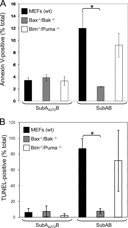
FIG. 2.
Quantitative analysis of SubAB-induced apoptosis in wt, bax−/−/bak−/−, and bim−/−/puma−/− MEFs. (A) In situ labeling of phosphatidylserine on the surface of apoptotic cells. Cells were treated with either SubAB (100 ng/ml) or SubAA272B (100 ng/ml) for 24 h. Phosphatidylserine was labeled with annexin V conjugated to Alexa 488 dye. (B) In situ TUNEL of DNA fragmentation in MEFs that were treated with either SubAB (100 ng/ml) or SubAA272B (100 ng/ml) for 30 h. Data represent the means ± SDs from two independent experiments. *, P < 0.05, Student's unpaired, two-tailed t test.

