FIG. 1.
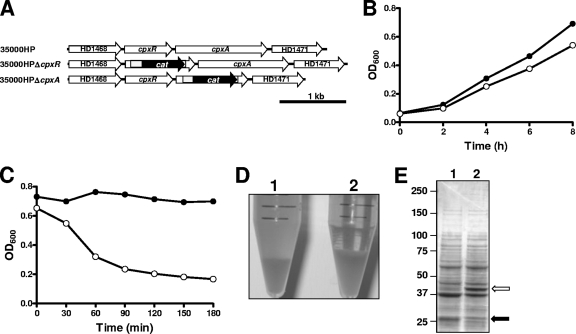
Characterization of the H. ducreyi cpxA deletion mutant. (A) Schematic of the cpxRA locus in the wild-type strain 35000HP, the 35000HPΔcpxR deletion mutant (35), and the 35000HPΔcpxA deletion mutant. (B) Growth of the wild-type parent strain 35000HP (closed circles) and the cpxA deletion mutant (open circles) in broth. Results from a representative experiment are shown. (C) Autoagglutination rates for the wild-type parent strain 35000HP (closed circles) and the cpxA deletion mutant (open circles). (D) Photograph of suspensions of the wild-type parent strain 35000HP (1) and the cpxA deletion mutant (2) after 3 h (at the end of the autoagglutination assay for which results are presented in panel C). (E) Total cell protein profiles for the wild-type parent strain 35000HP (lane 1) and the cpxA deletion mutant (lane 2) as determined by resolving proteins by SDS-PAGE and staining with Coomassie blue. The white arrow indicates the OmpP2B protein, which is overexpressed in the mutant, and the black arrow indicates the DsrA protein, which is present in the wild-type strain and missing in the mutant.

