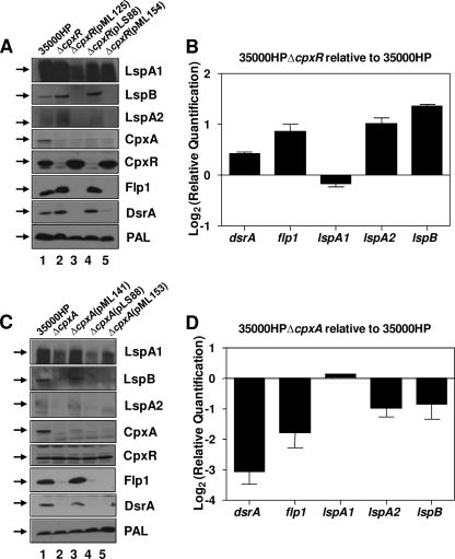
FIG. 2.
Deletion of cpxA or cpxR results in dysregulation of expression of several H. ducreyi gene products involved in virulence. H. ducreyi strains grown for 8 h in CB were used for preparation of whole-cell lysates or for RNA extraction. (A) Western blot analysis of whole-cell lysates of 35000HP (lane 1), 35000HPΔcpxR (lane 2), 35000HPΔcpxR(pML125) (lane 3), 35000HPΔcpxR(pLS88) (lane 4), and 35000HPΔcpxR(pML154) (lane 5); primary antibodies are indicated to the right of each panel. The PAL monoclonal antibody 3B9 (56) was used as a loading control for both panels A and C. It should be noted here that both LspA1 and LspA2 typically appear diffuse or exhibit a multiple banding pattern in Western blot analysis (35, 61). Black arrows on the left in panels A and C indicate the position of the relevant antigen. (B) Real-time RT-PCR showing transcript levels for dsrA, flp1, lspA1, lspA2, and lspB in 35000HPΔcpxR relative to 35000HP. (C) Western blot analysis of 35000HP (lane 1), 35000HPΔcpxA (lane 2), 35000HPΔcpxA(pML141) (lane 3), 35000HPΔcpxA(pLS88) (lane 4), and 35000HPΔcpxA(pML153) (lane 5); primary antibodies are indicated to the right of each panel. (D) Real-time RT-PCR showing transcript levels for dsrA, flp1, lspA1, lspA2, and lspB in 35000HPΔcpxA relative to 35000HP.

