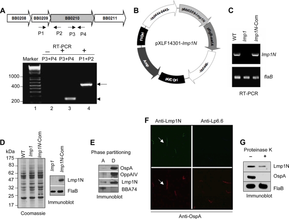
FIG. 7.
Genetic restoration of the lmp1N gene in the lmp1 deletion mutant. (A) lmp1 (bb0210) is cotranscribed with bb0209. The upper panel indicates schematic representation of the bb0209-bb0210 locus in the B. burgdorferi genome. The arrows indicate the positions of primer pairs used in RT-PCR analysis. The lower panel indicates RT-PCR analysis of cotranscription of bb0210 (lmp1) and bb0209. The DNA ladder in base pairs is noted to the left (lane 1). RT-PCR analysis in the absence (−; lane 2) or presence (+, lanes 3 and 4) of reverse transcriptase is shown. A 150-bp portion of bb0210 (lmp1) transcript (arrowhead) was amplified using primers P3 and P4 (lane 3), while a 520-bp portion of bicistronic transcript (arrow) spanning bb0209-bb0210 was amplified using primers P1 and P2 (lane 4). (B) Construction of pXLF14301-lmp1N. The putative promoter and open reading frame of lmp1 encoding Lmp1N and the streptomycin resistance cassette (flgBp-aadA) were cloned into pXLF14301 for recombination into B. burgdorferi chromosome. (C) Detection of the lmp1 gene expressing the lmp1N. Total RNA was isolated from the wild type (WT), lmp1 mutant (lmp1), or lmp1N-complemented (lmp1N-Com) B. burgdorferi strain, converted to cDNA, subjected to PCR analysis with flaB and lmp1 primers, and analyzed on a 1.5% agarose gel. (D) Detection of the Lmp1N protein. Lysates of B. burgdorferi were separated on an SDS-PAGE gel and were either stained with Coomassie blue (left) or transferred to nitrocellulose membrane and blotted with antiserum against Lmp1N or FlaB (right). (E) Triton X-114 phase partitioning of lmp1N-complemented B. burgdorferi proteins. Separated aqueous (lane A)- and detergent (lane D)-phase proteins were immunoblotted using Lmp1N antibody or antibodies against control hydrophilic (BBA74) or amphiphilic (OspA and OppA IV) proteins. (F) Immunofluorescence analysis. Unfixed lmp1N-complemented B. burgdorferi spirochetes were probed with antisera against Lmp1N or Lp6.6 and OspA followed by secondary antibodies, as detailed in Fig. 3B. The images were acquired with the 40× objective of a laser confocal microscope. (G) Lmp1N is surface exposed in the lmp1N-complemented isolate. The lmp1N-complemented B. burgdorferi strain was incubated in the presence (+) or absence (−) of proteinase K and subjected to immunoblotting with antisera against Lmp1N, FlaB, and OspA.

