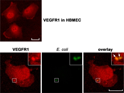
FIG. 3.
Colocalization of E. coli K1 with VEGFR1 in HBMEC. HBMEC cultured on coverslips were left untreated (top) or incubated with E44 for 15 min (bottom), and then the cells were fixed, permeabilized, subsequently stained with rabbit anti-VEGFR1 antibody, and incubated with Alexa Fluor 594-conjugated anti-rabbit IgG. The bacteria were stained with FITC-conjugated E. coli-specific antibody. Samples were analyzed using confocal laser scanning microscopy. The arrows indicate colocalization (yellow) of VEGFR1 and E. coli. Scale bar = 20 μm.

