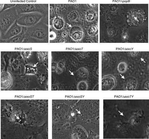
FIG. 1.
Phase-contrast microscopy images of human corneal epithelial cells at 8 h postinoculation with P. aeruginosa strain PAO1 or different T3SS mutants (Table 1). The 8-h window consisted of 3 h of incubation with 2 × 10⁷ CFU bacteria followed by 5 h of gentamicin treatment. Uninfected control cells are also shown. Arrows indicate the intracellular locations of P. aeruginosa within membrane bleb niches for strain PAO1 and other mutants which could express (and translocate) ExoS or within small intracellular vacuoles for strains with mutations in exoS (or which cannot translocate effectors, i.e., PAO1 ΔpopB). See Movies S1 to S3 in the supplemental material for a display of real-time microscopy images corresponding to wild-type PAO1 and effector mutants.

