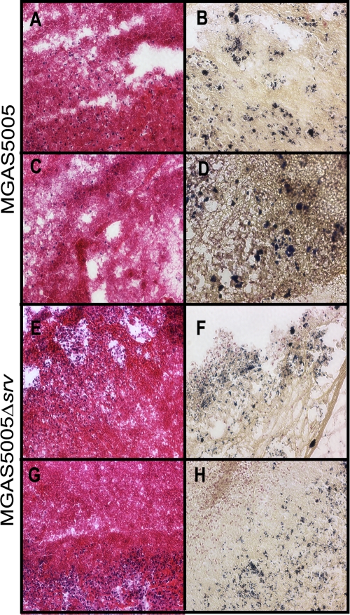
FIG. 6.
H&E staining (A, C, E, and G) and Gram staining (B, D, F, and H) of macroscopic structures removed at 7 dpi from the chinchilla middle ear cavity. MGAS5005 is represented in the top two rows, and MGAS5005 Δsrv is represented in the bottom two rows. H&E staining allows detection of host fibrin and polymorphonuclear leukocyte infiltration, whereas Gram staining allows detection of the Gram-staining phenotype, shape, and groupings of the bacteria. Magnification, ×20. Note the abundant microcolonies present in the MGAS5005 Gram-stained samples (B and D), whereas the bacteria appear more dispersed in the MGAS5005 Δsrv Gram-stained samples (F and H).

