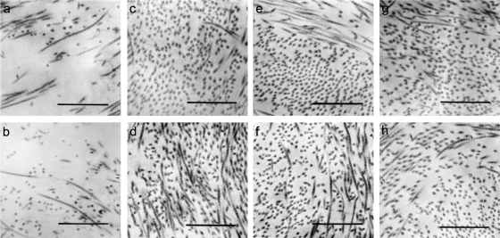
FIG. 4.
Plasma from placental blood stimulated with GBS induced extracellular matrix degradation in the amniochorion. Micrographs of the amniotic compact layer of the explants exposed to 10% plasma from placental blood stimulated with live (a) or heat-inactivated (b) GBS exhibit a drastic loss of collagen fibrils with amorphous material between them after 12 h of treatment. These alterations were inhibited by the addition of 0.1 μg/ml TIMP-1 (h), an MMP-9 inhibitor. Collagen fibrils appeared normally organized in bundles with specific orientation in explants treated with plasma from placental blood stimulated with live (c) or heat-inactivated (d) U. urealyticum and live (e) or heat-inactivated (f) L. acidophilus, as well as in control explants (g). Magnification, ×60,000. Bars, 0.0332 μm.

