FIG. 5.
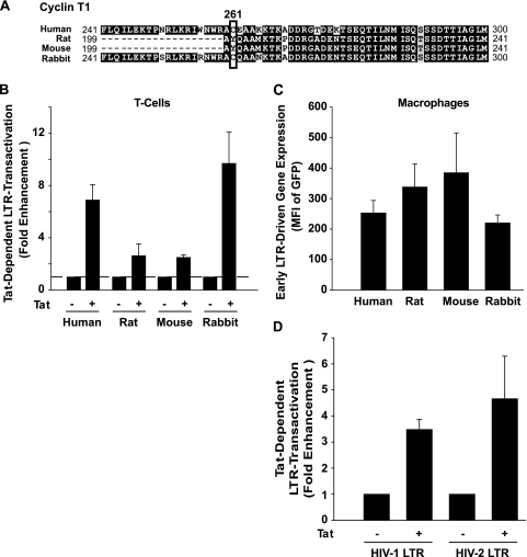
Tat-dependent HIV-1-LTR transactivation is species and cell type specific and occurs efficiently in primary rabbit cells. (A) Partial amino acid sequence alignment of cyclin T1s of human, rabbit, rat, and mouse origins (ClustalW2 method and BOXSHADE). Identical amino acids are shaded in black, conserved or similar residues are in gray, and unrelated amino acids are in white. Dashes indicate gaps introduced to optimize the alignment. Amino acid 261 is boxed. (B) Primary T cells were nucleofected with pHIV-1 LTR GFP in the presence or absence of an HIV-1 Tat expression vector. The MFI of GFP expression was quantified in viable cells 24 h later by flow cytometry. Shown are the arithmetic means ± SEM of results of duplicates or triplicates of two to four independent experiments. (C) Macrophages were infected with VSV-G HIV-1NL4-3 GFP and analyzed for Tat- and LTR-dependent reporter gene expression by flow cytometry 3 days later. Infectious titers were chosen such that the percentage of GFP-positive macrophages for all species was in the single-cell infection range (human, 1.0%; rat, 3.6%; mouse, 7.8%; rabbit, 0.4%). Shown are the arithmetic means ± SEM of results of duplicates or quadruplicates of two to four independent experiments. (D) Primary rabbit macrophages were transfected with pHIV-1 or pHIV-2 LTR GFP vectors in the presence or absence of an HIV-1 or HIV-2 Tat expression vector, respectively. The MFI of GFP expression was quantified 3 days later by flow cytometry. Shown are the arithmetic means ± SEM of results from two independent experiments.

