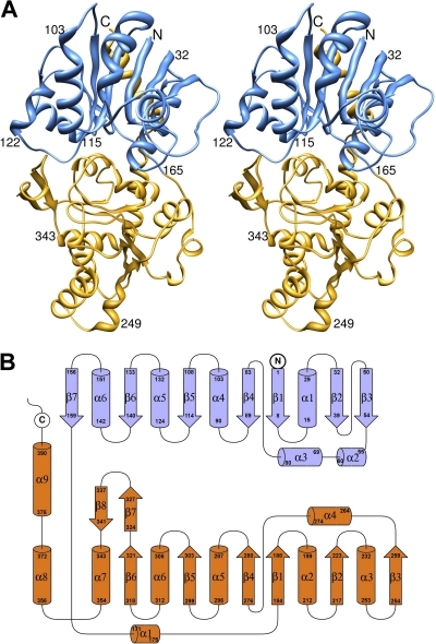
FIG. 2.
Crystal structure of glycosyltransferase expressed from virus NY-2A gene b736l. (A) Stereoribbon diagram of the B736L structure. The N- and C-terminal domains are colored blue and yellow, respectively. (B) Topology diagram of the B736L structure. The N- and C-terminal domains are colored blue and orange, respectively.

