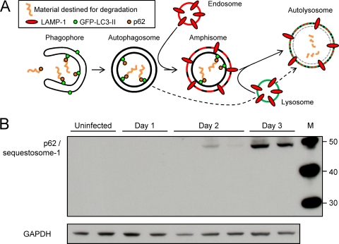
FIG. 2.
Accumulation of p62 indicates blockade of autophagic flux in CVB3-infected acinar cells in vivo. (A) The autophagy pathway is shown in diagrammatic form. (B) Western blotting was carried out to determine the amount of p62/sequestosome-1 in the pancreas at the indicated times p.i. GAPDH is used as a gel loading control. Each lane represents an individual mouse. A p62-specific antibody (from BD Biosciences; see Materials and Methods) was used to generate the data shown. Data are representative of those from two independent experiments. Similar results (data not shown) were obtained using a different p62-specific antibody (from Progen Biotechnik; see Materials and Methods).

