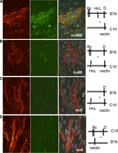
FIG. 1.
Fusion occurs when gB and gH/gL are in trans. (A to D) Nectin-1-expressing B78H1-C10 cells and/or B78 cells were transfected for 8 h with plasmids for the glycoproteins as shown in the stick diagrams. The C10 cells were trypsinized and overlaid onto coverslips containing B78 cells, and the bilayer was cocultured for 40 h. Cells were fixed and stained with anti-gH/gL polyclonal antibody (red). Propidium iodide was added to visualize nuclei (gray). Cells were analyzed by immunofluorescent assay for protein (red channel), EYFP (green channel), and nuclei (far-red channel). Images in the far-red channel were artificially colored white, as seen in the merged images (third set of images). Confocal images are at ×60 magnification and were captured using the same camera setting. n, number of syncytia per coverslip.

