FIG. 2.
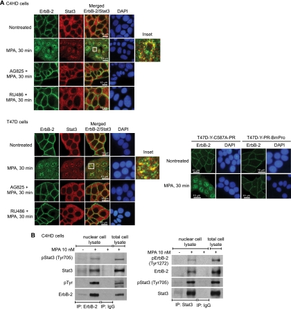
MPA induces Stat3 and ErbB-2 nuclear colocalization and physical association. (A) Cells were treated with MPA or pretreated with AG825 and RU486 before MPA stimulation. ErbB-2 (green) and Stat3 (red) were localized by immunofluorescence and confocal microscopy (see Materials and Methods for antibody specifications). Merged images in the third panels of the second rows show MPA-induced ErB-2 and Stat3 nuclear colocalization, evidenced by the yellow foci. The boxed areas are shown in detail in the right inset. (Bottom) T47D-Y cells were transfected with either PR-BmPro or C587A-PR mutants and were then treated with MPA. ErbB-2 (green) was localized by immunofluorescence as described above. Nuclei were stained with 4′,6-diamidino-2-phenylindole (DAPI) (blue). (B) Nuclear extracts from C4HD cells treated and untreated with MPA for 30 min were immunoprecipitated (IP) with ErbB-2 or Stat3 antibodies and analyzed by Western blotting with the indicated phosphotyrosine antibodies. Membranes were reprobed with total protein antibodies. As a control for the specificity of these protein interactions, lysates were immunoprecipitated with rabbit immunoglobulin G (IgG). Total cell lysates were blotted in parallel. The experiments from which the results were obtained were repeated three times, with similar results.

