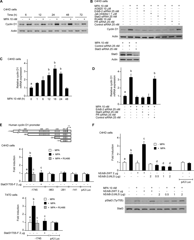
FIG. 4.
ErbB-2 acts as a Stat3 coactivator in MPA-induced cyclin D1 promoter activation. MPA induces cyclin D1 expression at the protein and mRNA levels via ErbB-2 and Stat3. (A) Cyclin D1 protein expression was analyzed by Western blotting. (B) Cells were preincubated with the indicated pharmacological inhibitors or transfected with Stat3, ErbB-2, and PR siRNAs and were then treated with MPA for 48 h. Cyclin D1 levels were studied by Western blotting. (Bottom) Control of inhibition of Stat3 expression by siRNAs (data not shown). Experiments from which the results in A and B were obtained were repeated three times, with similar results. (C) Cyclin D1 mRNA expression levels were determined by RT-qPCR. The fold change of mRNA expression levels upon MPA treatment for the indicated times was calculated by normalizing the absolute levels of cyclin D1 mRNA to GAPDH levels, which were used as an internal control, and setting the value of untreated cells as 1. (D) C4HD cells were transfected with Stat3, ErbB-2, PR, and control siRNAs and were then treated with MPA for 18 h. Cyclin D1 mRNA levels were studied by RT-qPCR, and data analyses were performed as described above for C. Data shown in C and D represent the means of data from three independent experiments ± standard errors of the means (SEM) (P < 0.001 for b versus a). (E) MPA induces cyclin D1 promoter activation via Stat3. Cells were transfected with a 1,745-bp-length human cyclin D1 promoter luciferase construct containing the GAS sites indicated at the top. C4HD cells were also transfected with constructs truncated at positions −963, −262, and −141, as shown in the diagram. When indicated, cells were cotransfected with the Stat3Y705-F expression vector. After transfection, cells were treated with MPA for 24 h. Results are presented as the fold induction of luciferase activity with respect to control cells not treated with MPA. The data shown represent the means of data from six independent experiments for each cell type ± SEM (P < 0.001 for b versus a and for c versus b). (F) ErbB-2 acts as a Stat3 coactivator. (Top) C4HD cells were transfected with the 1,745-bp cyclin D1 promoter construct as described above for E and were also cotransfected with the hErbB-2WT or hErbB-2ΔNLS vector when indicated and treated with MPA as described above for E. The relative light units of luciferase obtained in the transient transfection assays were normalized by the arbitrary densitometric values of phosho-Tyr 705/total Stat3 obtained in the Western blot shown at the bottom, and data are presented as the fold induction of cyclin D1 promoter activity relative to cells not treated with MPA. Data shown represent the means of data from three independent experiments ± SEM (P < 0.001 for b versus a, c versus b, and d versus b). (Bottom) Cells were transfected with hErbB-2WT or hErbB-2ΔNLS and were then treated with MPA for 10 min. Stat3 phosphorylation was studied by Western blotting as described in the legend of Fig. 1E (data not shown).

