Figure 7.
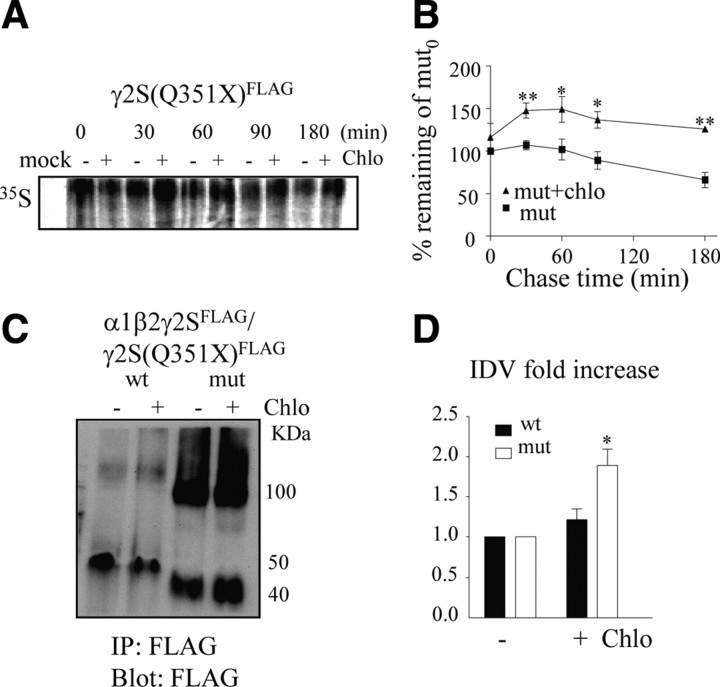
Mutant γ2S(Q351X) subunits were degraded through the lysosome pathway. A, Cells mock transfected (mock) or transfected with γ2S(Q351X)FLAG subunits were labeled with [35S]methionine for 20 min, followed by chase in the absence (−) or presence (+) of chloroquine (Chlo, 100 μm) for the indicated time periods. The cells were lysed, immunopurified using anti-Flag antibody, and analyzed by SDS-PAGE. B, The percentage radioactivity relative to the amount of radioactivity measured at time 0 for mutant subunits without chloroquine treatment in the absence (−) or presence (+) of chloroquine was plotted (*p < 0.05, **p < 0.01 vs mutant without chloroquine; n = 4). C, HEK 293T cells expressing equimolar concentrations of wild-type (wt) and mutant (mut) α1β2γ2SFLAG subunits were incubated without (−) or with (+) chloroquine (100 μm) for 4 h. The total lysates were immunopurified and immunoblotted with rabbit polyclonal anti-FLAG antibody. D, The total γ2 subunit protein IDVs with chloroquine treatment were normalized to the untreated group (*p < 0.05 vs wild type + chloroquine; n = 4).

