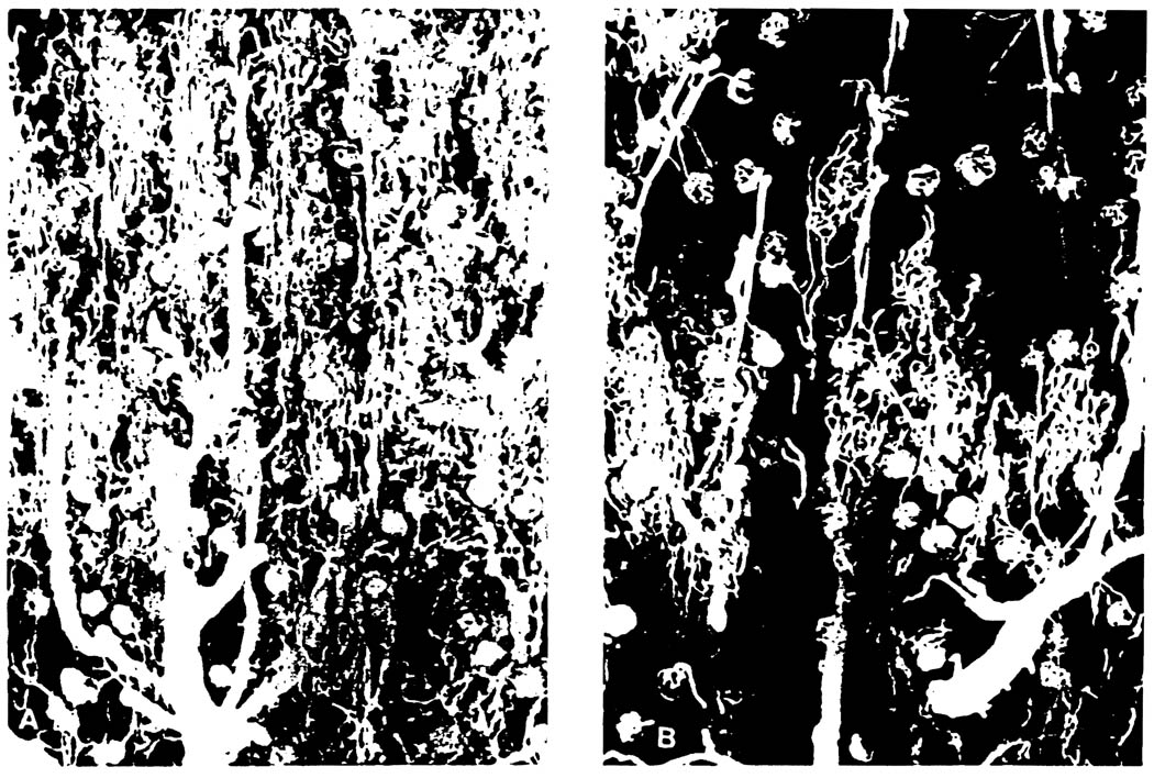
Fig 1.
Dissecting photomicrographs of renal cortical vasculatures visualized with silicon rubber compound at 1 hour after reperfusion of the graft (×50). (A) 72-hour EC kidney. Notice patchy distribution of avascular area, irregular and deformed pattern of interlobular artery and glomerulus. (B) 72-hour UW kidney. The glomerulus and capillary networks are fully filled with silicon rubber.

