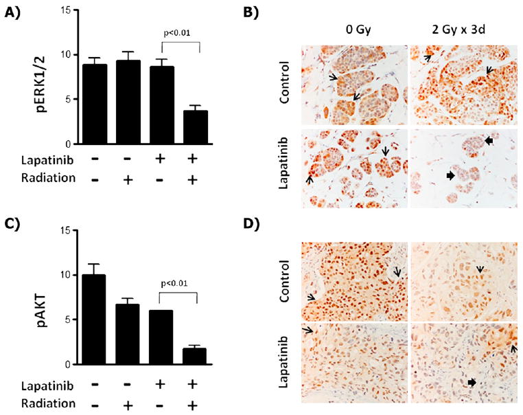
Fig. 3.

Radiosensitization by lapatinib correlates with inhibition of ERK1/2 in EGFR+/basal-like cells and with AKT in HER2+ breast cancer cells. (A) Tumors from basal-like/EGFR+ SUM149 xenografts were processed for immunohistochemistry with phosphorylated ERK1/2 antiserum and quantified from mice treated with lapatinib, radiotherapy, lapatinib plus radiotherapy, or vehicle control. (B) Sample immunohistochemistry staining of SUM149 tumors with phosphorylated ERK1/2 serum at 400×. Similarly, tumors from HER2+ SUM225 xenografts were processed for immunohistochemistry with phosphorylated AKT antiserum and (C) quantified. (D) Sample immunohistochemistry of SUM225 tumors with phosphorylated AKT serum at 400×. Open arrows indicate areas of increased staining; solid arrows, areas of reduced staining.
