
Figure 2.
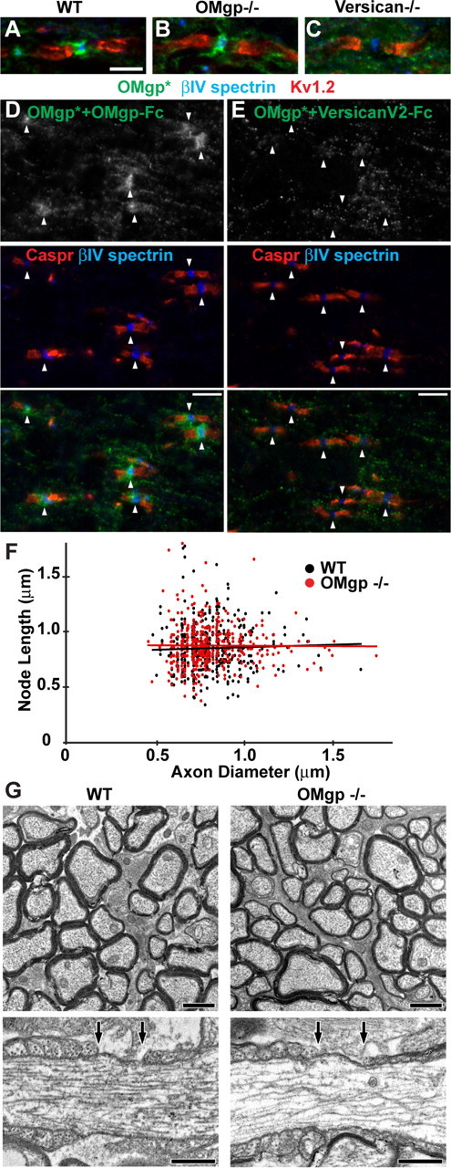
α-OMgp* detected versican V2 at CNS nodes of Ranvier. A–C, Optic nerve sections of WT, OMgp-null, and versican V0/V2-null mice stained with α-OMgp* (green), anti-βIV spectrin (blue), and anti-Kv1.2 (red). Scale bar, 5 μm. D, E, α-OMgp* was preadsorbed with OMgp-Fc (D) or versican V2-Fc (E) before being incubated with WT spinal cord sections. Arrowheads indicate nodes of Ranvier. Scale bars, 5 μm. F, Node length plotted as a function of axon diameter in WT and OMgp-null optic nerves. G, Electron micrographs of optic nerves from 100-d-old wild-type and OMgp null (−/−) mice. Top panels show cross sections of myelinated axons in the optic nerves. Lower panels show single nodes of Ranvier from longitudinal sections of optic nerves. The nodal gap is defined by the two arrows above each axon. Scale bars: top, 1 μm; bottom, 0.5 μm.