
Fig. 4. Two-color brightfield micrographs (panels A–F) from sections immunostained with antisera to Eps8 (blue) and Tbr2 (brown) show varying densities of UBCs in folia of lobules VII (CB-VII) and X (CB-X) of the cerebellar cortex and in regions of the CN. Panels G and H (two-color immunofluorescence) show individual UBCs from cerebellum CB) and DCN and illustrate cytoplasmic staining by Eps8 antiserum (red) and nuclear staining by Tbr2 antiserum (green).
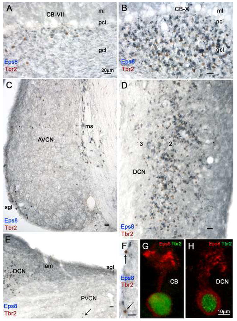
A and B: Even at this low magnification it is evident that the granular layers (gcl) of the two lobules contain different densities of intensely Eps8+ neuropil structures and Tbr2+ nuclei, identified at higher magnification as brushes and nuclei of UBCs, respectively (see panel G). Granule cell glomeruli and molecular layer (ml) parallel fibers are weakly Eps8 immunoreative; the Purkinje cell layer (pcl) is unstained. C: This coronal section of the AVCN demonstrates higher density of UBCs in the medial sheet (ms) than in the superficial granular layer (sgl) and the magnocellular AVCN; moreover, the density of UBCs is higher in rostral than ventral parts of AVCN. D: Detail from a coronal section tilted 90° shows high density of UBCs in layer 2 and the polymorphic layer 3 of the DCN, while granule cell glomeruli and molecular layer 1 parallel fibers are weakly Eps8+. E: The lamina (lam) shows moderate Eps8 staining of granule cells and glomeruli, while numerous UBCs are present in the adjacent DCN and one UBC (arrow) is present in the magnocellular PVCN. F: Two UBCs (arrows) and several granule cells (arrowhead) in the superficial granular layer of AVCN. G and H: UBCs in the granular layer of the cerebellum (CB) and DCN. Magnification bars A–F = 20 μm; G, H = 10 μm.