
Fig. 5. Morphological UBC variants in the CN demonstrated by laser confocal immunofluorescence (panels A, B, D, E, H, K, L, and J) and brightfield microscopy (panels C and I).
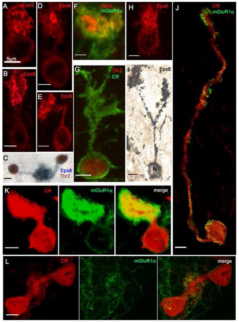
A: The bulbous brush of this UBC arises directly from the cell body and contains multiple, fragmented Eps8+ foci. B: The elongated brush of this UBC arises from a short and straight dendritic stem and contains variously shaped Eps8+ foci. C: Two UBCs, double-labeled with Eps8 and Tbr2, contribute brushes to the same glomerulus. D: UBC brush with large, ring-like Eps8+ focus. E: UBC with sharply bent dendritic stem. F: Detail of an UBC brush (identified by peripheral staining with mGluR1α) that contains two large Eps8+ foci. G: This CR+/Tbr2+ UBC shows a loose array of brush dendrioles. H: This UBC, stained with Eps8, emanates dendrioles from different parts of the cell body (compare with panel A). I: This UBC, stained with a silver-enhanced Eps8/DAB protocol, shows two brushes formed by branches of a long dendritic stem. J: CR+/mGluR1α+ UBC emits an unusually long dendrite that forms a small, elongated brush. Row K: This UBC has a very short dendrite and a large, compact brush; dual immunostaining of both cell body and brush indicates mixed, CR+/mGluR1α+ phenotype. Row L: The brushes of two CR+/mGluR1α− UBCs merge into the same glomerular array. Magnification bars = 5 μm.