
Figure 5.
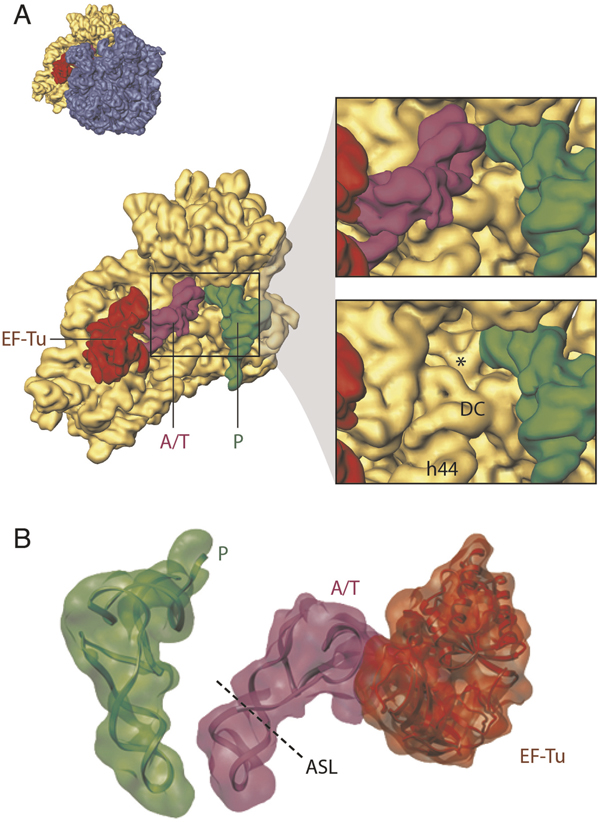
Decoding and aa-tRNA incorporation. (A) Close-up view of the 30S subunit showing the decoding centre (DC) region in the presence of a cognate ternary complex. In the lower panel, the tRNA is computationally removed to show the tip of helix 44, in which A1492 and A1493 flip out as the result of a cognate codon-anticodon interaction. The resulting configuration is labelled with an asterisk. The orientation of the small subunit is shown as successive thumbnails on the left. (B) Arrangement of the P-site tRNA and ternary complex. The tRNA atomic models (displayed in ribbons) were obtained by fitting of the experimental cryo-EM densities (shown as transparent) with the X-ray-derived coordinates of the tRNAs by real-space refinement.