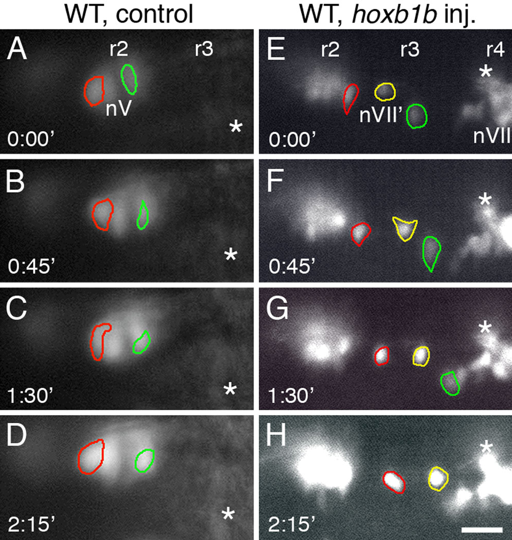
Figure 2.

FBMN-like neurons in r2 and r3 move along the anterior-posterior (AP) axis of the hindbrain. All panels show dorsal views of the hindbrain, with anterior to the left. Panels A–D and E–H show a sequence of images, taken at 45 minute intervals, of GFP-expressing neurons in the r2/r3 region (on one side of the hindbrain) in a control wild-type embryo (A–D) and in a wild-type embryo injected with hoxb1b RNA (E–H). Since the earliest GFP-expressing nV neurons in r3 of control embryos arise after 26 hpf (Higashijima et al., 2000), we compared behaviors of nV neurons in r2 of control embryos to nVII’ neurons in the broader r2/r3 region of hoxb1b-injected embryos. Particular cells have been outlined in different colors to monitor their positions over the 135-minute observation period. Fiducial marks (asterisks) are indicated in successive frames. (A–D) In the control embryo, the two nV neurons (red and green outlines) in r2 change shape over time, but do not move along the AP axis. (E–H) In the hoxb1b-injected embryo, the FBMN-like cells (nVII'; red, green and yellow outlines) are located in r2 or r3, adjacent to a cluster of putative nV neurons in r2. The three cells change shape over time, but also move posteriorly, toward the cluster of FBMNs in r4 (nVII). Scale bar, 50 µm.
