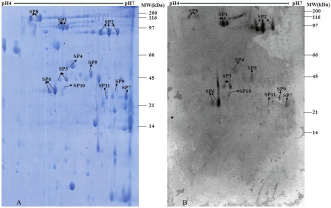
Figure 2. 2-D proteome reference map and representative immunoblot of ECPs of B.pertussis Chinese WCV strain 58003.
ECPs were separated by IEF at pH 4–7 in the first dimension and then by 12.5% SDS-PAGE in the second dimension. Gels were either Coomassie blue-stained (Fig. 2A) or immunoblotted with a 1∶1000 dilution of pooled immune sera from vaccinated children (Fig. 2B). The protein spots of interest were excised individually for identification by PMF. The spot numbers refer to the identified immunoreactive proteins listed in Table S1.

