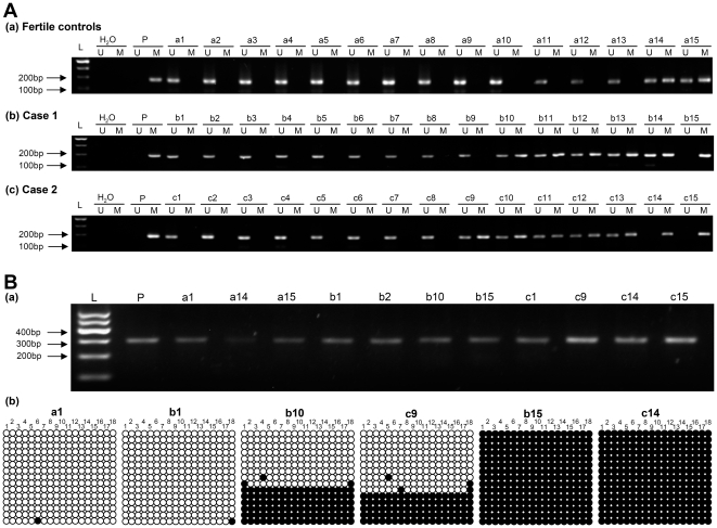
Figure 3. Methylation status of the promoter of MTHFR in genomic DNA prepared from ejaculated human sperm.
(A) Representative results of the MSP analysis of MTHFR. DNA obtained from the sperms was amplified with primers specific to the unmethylated (U) or the methylated (M) of MTHFR after treatment with sodium bisulfite. (a) Fertile controls a1–a13 showed only the unmethylated allele. Others showed both methylated and unmethylated alleles. (b) Idiopathic infertile males with normozoospermia (Case 1) b1–b9 showed only the unmethylated allele. Idiopathic infertile males with normozoospermia b10–b14 showed both methylated and unmethylated alleles. Other showed only the methylated allele. (c) Idiopathic infertile males with oligozoospermia (Case 2) c1–c8 showed only the unmethylated allele. Idiopathic infertile males with oligozoospermia c9–c13 showed both methylated and unmethylated alleles. Others showed only the methylated allele. (B) Representative results of bisulfite-PCR sequencing of MTHFR. Filled and open circles represent methylated and unmethylated CpGs, respectively. L: molecular weight markers; P: positive control (universal methylated DNA).

