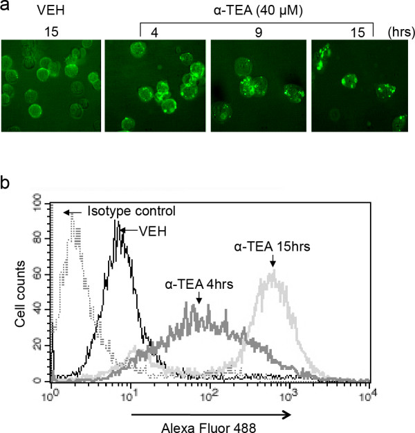
Figure 2.

α-TEA induced increased levels and aggregation of ceramide in cell surface membranes. A, MDA-MB-231 cells were treated with 40 μM α-TEA for 4, 9 and 15 hrs (15 hrs for vehicle control). The living cells were collected and immunostained with antibody to ceramide. Cellular membrane ceramide was assessed by fluorescence microscope. B, Samples from Figure 2a at 4 and 15 hrs time points were also assessed by FACS for membrane expression of ceramide. All images are representative of three independent experiments.
