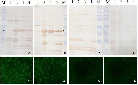
Figure 3.

Western-blotting analysis of the expressed recombinant GST-ORF2-E protein with porcine serum (above) was confirmed by IFA (below). A clear band with the expected molecular weight appeared on the NC membrane after incubation with two positive porcine serum samples (A, B), but no equal band appeared when incubated in two samples of negative porcine serum (C, D). Lane 1, BL21 cell lysate before induction of IPTG; Lane 2, BL21 cell lysate after induction of IPTG; Lane 3, Supernatant of cell lysate after sonication and centrifugation; Lane 4, Pellet of cell lysate after sonication and centrifugation; Protein marker includes 8 bands of 175, 83, 62, 47.5, 32.5, 25, 16.5, and 6.5 kDa.
