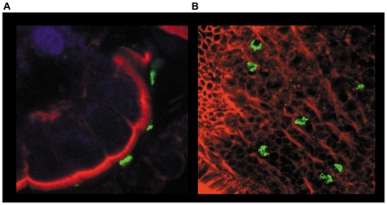
Figure 1. Ex-vivo colonisation of human intestine by C. jejuni.
Human intestinal biopsies from the terminal ileum were co-cultured for 12 hrs with WT C. jejuni 11168H. Following co-culture, bacteria were localized by immuno-labelling with primary unlabelled anti-campylobacter antibody and secondary FITC labelled antibody (green). Actin filaments including apical brush border were visualized with rhodamine phalloidin (Red). TO-PRO blue was used to counter-stain for nuclei (blue). Whole tissue samples were examined by confocal microscopy. Transverse cross-section of the tissue (a) and an apical view (b) are shown.

