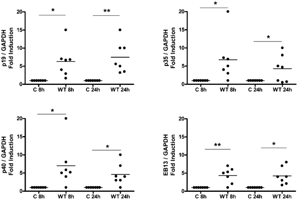
Figure 3. Monocyte-derived DC (DC) cytokine milieu in response to C. jejuni 11168H wild-type strain.
DCs incubated in media alone served as Control (C) or were infected with C. jejuni 11168H wild-type (WT) strain (multiplicity of infection; MOI = 100). mRNA expression of the IL-12 family members (p19, p35, p40, EBI3 at 8 and 24 hours) was quantified by RT-PCR. (a) Gene expression was normalised to GAPDH. Variations in mRNA levels are expressed as fold induction compared to the uninfected control cells. (Median is shown). A representative gel to highlight variation in subunit expression between donors (D) is included (see Figure S1).

