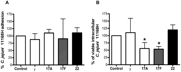
Figure 6. IL-17A and IL-17F reduce C. jejuni 11168H intracellular survival in intestinal epithelia.
Confluent Caco-2 cells were exposed to individual cytokines for 24 hours prior to infection with C. jejuni 11168H WT strain (MOI = 100) for 3 hours at 37°C. Cell lysates were serially diluted and plated for total viable bacterial counts (adhesion + invasion). In parallel, another set of infected cells was exposed to 150 µg/ml gentamicin for 2 hours (to kill extracellular adhered bacteria) and lysates plated for enumeration of viable intracellular bacteria. Data represent average percentage cfu obtained in treated versus untreated cells (the latter set at 100%). Statistical analysis of 3 independent experiments performed in duplicate is shown versus untreated cells (Data shown as the median + range).

