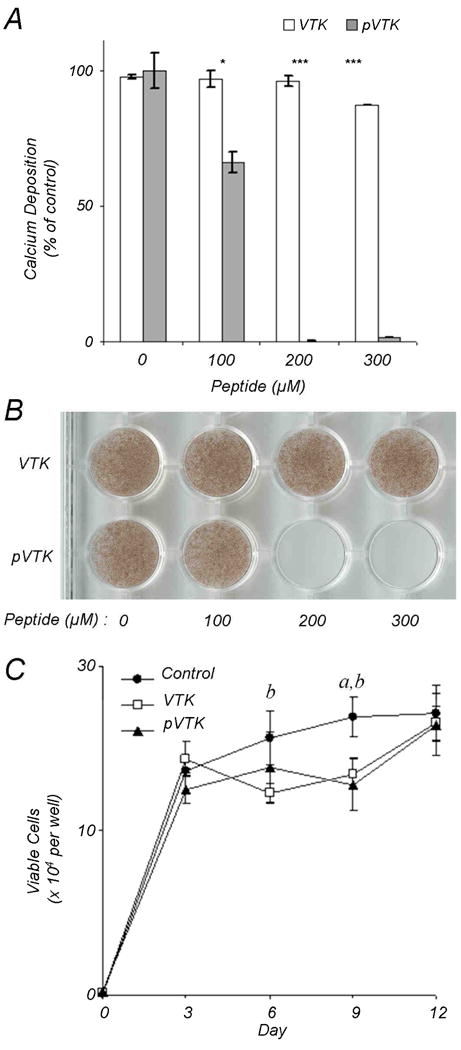
Fig. 2.

Mineralizing MC3T3-E1 osteoblast cultures were incubated with VTK and pVTK at the indicated concentrations for 12 days followed by mineral quantification by (A) calcium content determination expressed as a percentage of untreated control cultures, and (B) von Kossa (silver nitrate) staining for mineral. Data are presented as means ± S.D. *p < 0.05; ***p < 0.001 from Student's t-test for statistical differences between the two peptides at a given dose. (C) Cell proliferation in osteoblast cultures treated with, or without, 200 μM pVTK and VTK as measured by MTT assay. a denotes statistical significance between Control and VTK at the given time point. b denotes statistical significance between Control and pVTK at the given time point.
