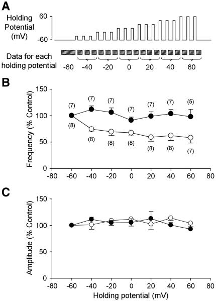
Figure 5.
Effects of postsynaptic membrane depolarization on presynaptic GABA release. A. Depolarization-induced disinhibition protocol. Brief (20 s) depolarizing steps were applied to the postsynaptic neurons every minute. Two minutes of mIPSC recordings before the first depolarizing step was used for the −60 mV data. Each voltage step was applied three times and the mIPSCs after each depolarizing step were analyzed for the data for each step. B. Group data of mIPSC frequency at different postsynaptic membrane potentials in the absence (open circles) and presence (closed circles) of the CB1R antagonist, AM 251 (5 μM). Postsynaptic depolarization significantly decreased mIPSC frequency, but not amplitude, suggesting a decrease in presynaptic GABA release (p<0.05 for postsynaptic membrane potential and interaction, two-way ANOVA.). C. Group data for mIPSC amplitude at different postsynaptic membrane potentials in the absence (open circles) and presence (closed circles) of the CB1R antagonist. Numbers in parentheses indicate number of neurons.

