
Figure 5.
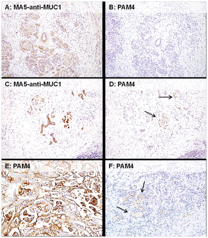
Immunohistology of tissue specimens derived from patients with primary diagnoses of chronic pancreatitis and pancreatic adenocarcinoma. The upper panels (A & B) are from a single patient specimen and are representative of 90% of the pancreatitis specimens (18 of 20 evaluable). The MA5 anti-MUC1 (peptide core) antibody, employed as a positive control, was reactive with acinar, ductal and ADM cells (A), whereas the PAM4 antibody was negative for all cell types (B). The middle panels (C & D) are from a single patient specimen where the MA5 antibody gave an intense and diffuse labeling of the acinar, ductal and ADM cells (C), whereas the PAM4 antibody gave only a focal, weak reactivity with ADM (D). The bottom panels (E & F) are two individual specimens of pancreatic adenocarcinoma, each labeled with PAM4 antibody. Panel E, representative of the majority of pancreatic adenocarcinomas, shows an intense, diffuse labeling of adenocarcinoma cells and secreted mucin, whereas panel F shows a weak, focal labeling of the adenocarcinoma cells. The arrows in panels D and F point to tissues that are weakly labeled with PAM4. (200x for all magnifications)