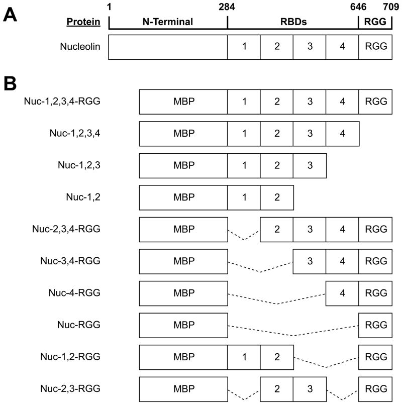
Fig. 2.
Nucleolin deletion mutants. (A) Diagram of nucleolin structure. (B) Diagram of the nucleolin deletion mutants used in this study. Solid lines indicate regions of the nucleolin peptide that have been deleted from the Nuc-1,2,3,4-RGG construct. All proteins were overexpressed in E. coli fused at the N-terminal to the maltose-binding protein (MBP).

