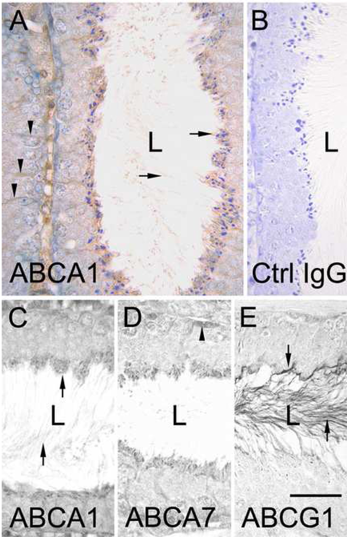
Figure 1. Immunolocalization of ABCA1, ABCA7 and ABCG1 in mouse seminiferous tubules.
A, anti-ABCA1 immunohistochemical labeling of a seminiferous tubule at stage VII of the cycle. Arrows in A points to staining of the heads of late step 16 spermatids (located at apical border) and on the tails of the spermatids in the lumen (L). Arrowheads in A point to anti-ABCA1 staining in Sertoli cells. B, control IgG labeling of a seminiferous tubule. C, D and E, comparative immunolabeling of seminiferous tubules at stage VII using antibodies to ABCA1, ABCA7 and ABCG1, respectively. Arrows in C point to ABCA1 staining of the heads and tails of step 16 spermatids. Arrowheads in D point to anti-ABCA7 staining of a spermatogonium. Arrows in E point to anti-ABCG1 staining of the head and tail (in lumen) of late step 16 spermatids. Bar in A=30µm and applies to A and B. Bar in E=25µm and applies to C–E.

