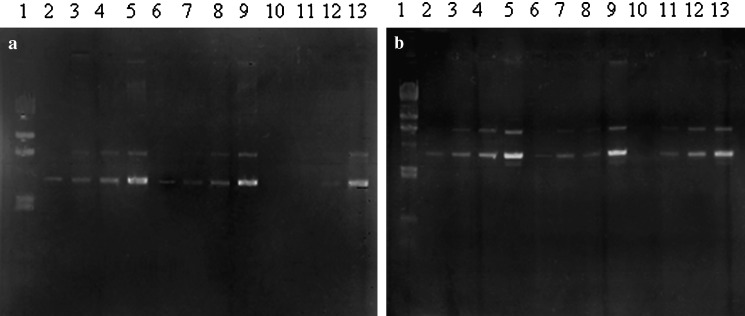
Fig. 2.
Agarose gel electrophoresis of a pDNA-loaded Ca-pectinate, b pDNA-loaded Mg-pectinate. Lane1 Marker; lanes 2–5 0.01, 0.05, 0.1, and 0.5 μg of pDNA, respectively; lanes 6–9 pectin/CaCl2 = 0.1:0.5 or pectin/MgCl2 = 0.5:50 with 0.01, 0.05, 0.1, and 0.5 μg of pDNA, respectively; lanes 10–13 pectin/CaCl2 = 0.2:1.0 or pectin/MgCl2 = 1:100 with 0.01, 0.05, 0.1, and 0.5 μg of pDNA, respectively

