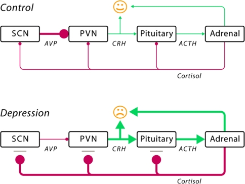
Fig. 1.
Schematic illustration of the pathogenesis of depression (reprinted with permission from Bao et al., 2008 [13], p. 541). The schematic figure illustrates the impaired interaction among the decreased activity of vasopressin neurons (AVP) in the suprachiasmatic nucleus (SCN), the increased activity of corticotropin-releasing hormone (CRH) neurons in the paraventricular nucleus (PVN), the increased release of adrenocorticotropin (ACTH) into the blood stream by the pituitary gland, and the increased release of cortisol by the adrenal gland. Normally, cortisol exerts a negative feedback effect to shut down the stress response when the threat has passed. In depressed patients, the cortisol feedback mechanism is deficient due to the presence of glucocorticoid resistance, which may be caused either by polymorphisms of the corticosteroid receptor or by early (intra-uterine or childhood) developmental disorders. Both, increased CRH and increased cortisol levels may induce mood disorders by their central effects. The increased cortisol levels also affect the vasopressin (AVP) neurons in the suprachiasmatic nucleus (SCN) as they subsequently fail to inhibit sufficiently the CRH neurons in the hypothalamic paraventricular nucleus (PVN). Such an impaired negative feedback mechanism may lead to a further increase in the activity of the HPA system [13]

