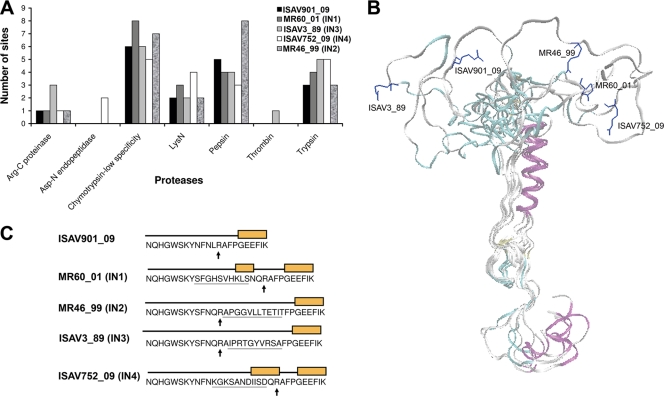
FIG. 3.
Homology modeling of fusion proteins of ISAV. (A) Number of sites for several proteases present in IN of fusion proteins. (B) Superposition of models of the ISAV fusion protein with different insertions, and putative site of the proteolytic processing site (residue R) (the amino acid residue is shown in a stick representation). The models were generated by homology modeling, using the fusion domain from the HA protein of influenza A virus as a template. (C) Putative secondary structure of IN. The boxes show the helix structure.

