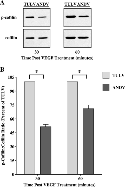
FIG. 5.
Decreased cofilin phosphorylation in ANDV-infected ECs following VEGF treatment. (A) Cofilin phosphorylation within ANDV-infected ECs following VEGF treatment was assessed by Western blotting. HUVECs were infected with ANDV or TULV (MOI of 0.5), and at 3 days p.i., cells were treated with VEGF for the indicated times. Equivalent amounts of total protein were separated by SDS-polyacrylamide (15%) gel electrophoresis and analyzed by Western blotting using anti-cofilin or anti-phospho-cofilin rabbit polyclonal antibodies (Cell Signaling), HRP-conjugated anti-rabbit secondary antibody (Amersham), and enhanced chemiluminescence (Amersham). Ninety percent of cells were infected by ANDV and TULV, respectively. (B) Densitometric analysis of cofilin and phospho-cofilin levels was performed using ImageJ (NIH) software, and data were plotted as the means ± SEM using GraphPad Prism 5 software. *, P < 0.005.

