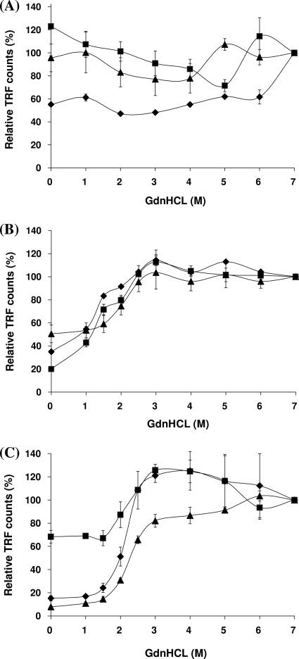
FIG. 1.
Change in CDI TRF counts following exposure to increasing concentrations of GdnHCl. Brain homogenates from the frontal cortex were treated with increasing concentrations of GdnHCl (range, 0 to 7 M) and measured by CDI. Three cases each from neurological control (A), vCJD (B), and sCJD subtype MM1 (C) were analyzed. Each symbol (squares, diamonds, and triangles) represents individual cases of each phenotype. TRF counts at particular concentrations of GdnHCl were expressed as relative values (percentages) to those at 7 M GdnHCl. Data shown represent the average values ± SD for triplicate wells except for one control case (diamonds in panel A, duplicate wells). The non-CJD cases presented were all of the PRNP codon 129 MM genotype.

