Fig. 2.

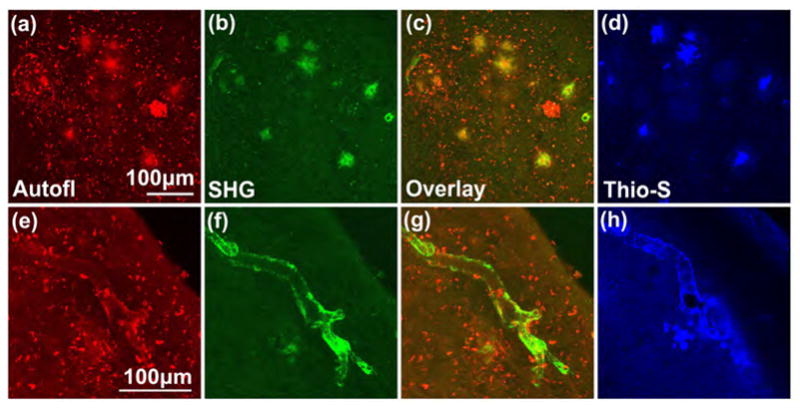
Senile plaques emit autofluorescence and second harmonic signal. (a) Autofluorescence and (b) second harmonic emissions detected in the entorhinal cortex in acute slices of a 22-month old APPSwe/TauJNPL3 mouse. (c) Overlay of (a) and (b) shows the same large, round structures (appears as yellow in the overlay) emit both the autofluorescence and second harmonic signals, which were then identified to be senile plaques when (d) this brain slice was subsequently fixed and stained with the plaque-specific dye Thioflavin-S. The small, bright specks were lipofuscins, which did not generate second harmonic emission. (e-h) Autofluorescence and second harmonic signal, of unknown molecular origin, were also detected near a blood vessel that was affected by cerebral amyloid angiopathy. Signal could be emitted from a source similar to that in senile plaque or from collagen. Each panel is a z-projection of a 40–60μm thick image stack acquired in 3 or 4μm steps. Multiphoton excitation wavelength = 774nm, circular polarization.
