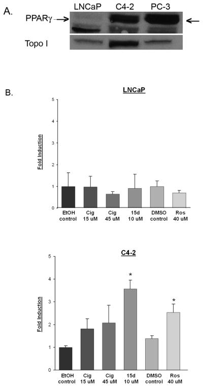
Fig 1. C4-2 cells express functional PPARγ.
(A) Nuclear extracts were prepared from the C4-2, LNCaP and PC-3 cell lines. The PPARγ protein present in each nuclear fraction was detected using Western blot analysis. The polyclonal PPARγ antibody used in these studies detects PPARγ (indicated by arrow) as well as a smaller, non- specific band. The blots were stripped and reprobed with an antibody against topoisomerase I (Topo I) to confirm purity of nuclear fractions. (B) LNCaP and C4-2 cells were transfected with the PPRE3-luciferase and CMV β-galactosidase plasmid constructs. Transfected cells were then treated with vehicle control (EtOH or DMSO) or PPARγ ligands at various concentrations for 24 hours. Luciferase activity was measured and normalized to β-galactosidase activity. Each bar represents the mean ± SD of three wells. *, P< 0.05 compared to vehicle control. A representative experiment is shown.

