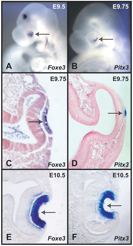
Figure 1.
In situ hybridizations of Foxe3 and Pitx3 probes to wild type mouse embryos. (A) Whole mount in situ hybridization of a Foxe3 probe to an E9.5 embryo showing expression in the lens placode (arrow). (B) Whole mount in situ hybridization of Pitx3 probe to an E9.75 embryo showing expression in a part of the lens placode (arrow). (C) A section showing the expression of Foxe3 in the lens placode (arrow). (D) A section through the embryo in B showing expression of Pitx3 in a few cells of the lens placode (arrow). (E) A section of an E10.5 embryo hybridized with Foxe3 probe showing strong expression in the lens pit (arrow). (E) A section of an E10.5 embryo hybridized with Pitx3 probe showing strong expression in most of the cells of the lens pit (arrow).

