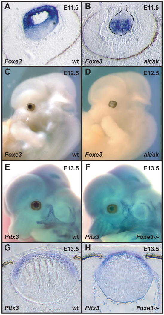
Figure 2.
In situ hybridizations of Foxe3 and Pitx3 to wild type, ak and Foxe3-deficient embryos. (A) A section of an E11.5 wild type embryo hybridized with Foxe3 probe showing strong labeling in the developing lens. (B) A section of an E11.5 ak embryo hybridized with a Foxe3 probe showing expression of this gene in the abnormal lens. (C) Whole mount in situ hybridization of Foxe3 probe to an E12.5 wild type embryo showing expression in the lens. (D) Whole mount in situ hybridization of Foxe3 probe to an E12.5 ak embryo showing dramatically reduced expression of this gene in the lens. (E) Whole mount in situ hybridization of Pix3 probe to an E13.5 embryo wild type embryo showing expression of this gene in the lens. (F) Whole mount in situ hybridization of Pitx3 probe to an E13.5 Foxe3-deficient embryo showing only slightly reduced levels of expression of this gene in the lens. (G) A section through the embryo in (E) showing the expression of Pitx3 in wild type embryo. (H) A section through the embryo in (F) showing the expression of Pitx3 in Foxe3-/- embryo.

