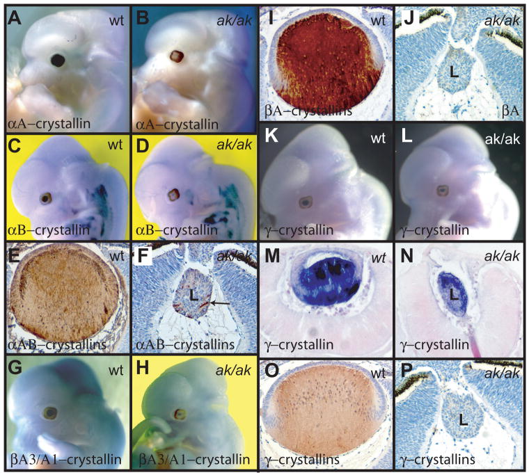
Figure 4.
Analysis of crystallin expression in E12.5 wild type and ak embryos. (A, B) Whole mount in situ hybridizations of an αA-crystallin (Crya1) probe to a wild type embryo (A) and to an ak embryo (B). (C, D) Whole mount in situ hybridizations of an αB-crystallin (Crya2) probe to a wild type embryo (C) and to an ak embryo (D). (E, F) Immunostaining of lens sections from a wild type embryo (E) and an ak embryo (F) with antibodies directed against αA and αB-crystallins. Only few cells show expression in the ak lens (arrow). (G, H) Whole mount in situ hybridizations of βA3/A1-crystallin (Cryba1) probe to a wild type embryo (G) and an ak embryo (H). (I, J) Immunostaining of lens sections from a wild type embryo (I) and an ak embryo (J) with antibodies directed against βA-crystallins. (K, L) Whole mount in situ hybridizations of γ-crystallin probe to a wild type embryo (K) and to an ak embryo (L). (M) A section of the embryo in (K) showing high levels of γ-crystallin expression in the posterior of the lens. (N) A section of the embryo in (L) showing γ-crystallin expression in the anterior part of the lens. (O, P) Immunostaining of lens sections from a wild type embryo (O) and an ak embryo (P) with antibodies direct against γ-crystallins.

