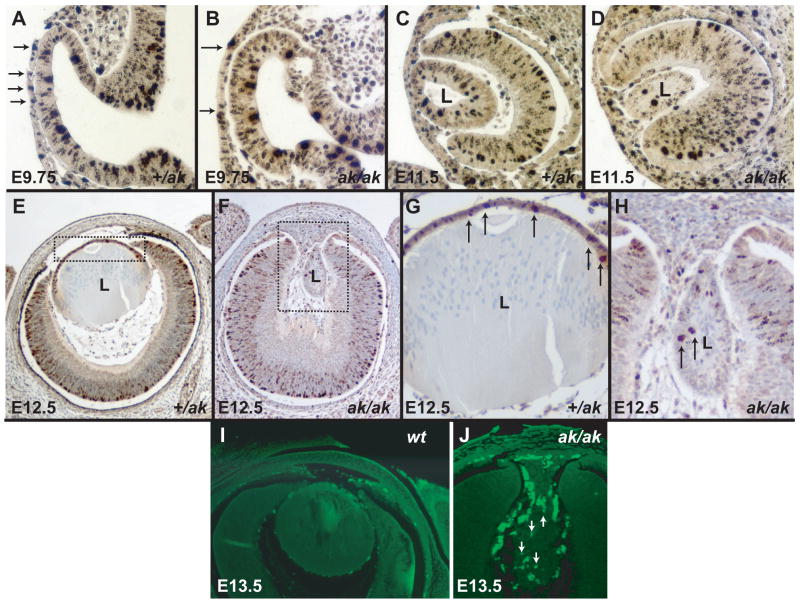
Figure 6.
Proliferation and apoptosis in Pitx3 heterozygous and Pitx3-deficient embryos. (A) A section of an E9.75 +/ak eye reacted with antibody against phosphohistone H3. Arrows point to the phH3 positive cells. (B) A section of an E9.75 ak/ak eye showing reaction with an antibody against phosphohistone H3. Arrows bracket the presumptive lens placode that shows a reduced labeling with antibodies against phH3. (C) A section of an E11.5 +/ak eye reacted with antibody against phosphohistone H3. (D) A section of an E11.5 ak/ak eye showing reaction with the antibodies against phosphohistone H3. (E) A section of an E12.5 +/ak eye reacted with antibody against phosphohistone H3. The boxed region is magnified in G. (F) A section of an E12.5 ak/ak eye showing reaction with the antibody against phosphohistone H3. The boxed region is magnified in H. Arrows indicate phosphohistone H3 positive cells. (I, J) TUNEL assay on the wild type eye (I) and on the ak eye (J). Apoptosis is not observed in the wild type lens, while TUNEL positive cells (fluorescent nuclei) are seen in the ak lens (arrows). Fluorescent cells surrounding the lens are likely to be of neural crest origin.

