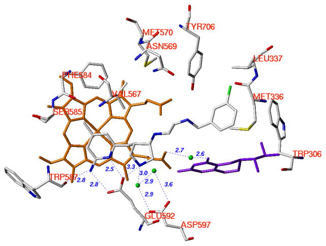
Figure 5.
Crystallographic binding conformations of 23 in complex with nNOS (PDB id: 3B3P).27 The heme (orange), H4B (purple), and structural water molecules (green) involved in the binding of 23 to nNOS are shown. The active site residues and ligands are represented in an atom-type style (carbons in grey, nitrogens in blue, oxygens in red, and sulfur in yellow). The distances of some important H-bonds between the residues, structural water molecules, cofactors, and inhibitors are given in Angstroms (Å).

