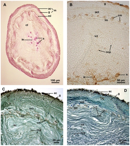
Figure 1. Histochemical labeling of the Cuvierian tubules of Holothuria forskali.
(A, C, D: paraffin sections; B: cryo-section). A: Cross section through a whole tubule stained with the PAS method; B–D: Transverse sections through the tubule wall labeled with PSA, Con A and LCA, respectively (C and D were counterstained). ac: adluminal cell layer of the mesothelium; cm: circular muscle; ct: connective tissue; g: granular cell layer of the mesothelium; ict: inner connective tissue layer; ie: inner epithelium; lm: longitudinal muscle; m: mesothelium; ml: muscular layer; ncp: neurosecretory-like cell processes; oct: outer connective tissue layer; s: spherulocytes.

