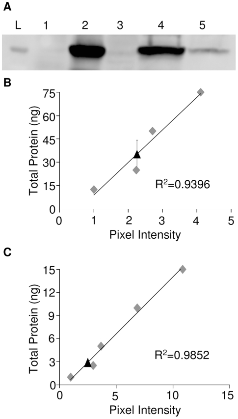
Figure 4. Cap- and IRES- dependent GFP expression levels in mosquito cells.
(A) Detection of GFP by Western blot in uninfected C6/36 cells (lane 1), C6/36 cells infected with dsSINV/GFP (lane 2), dsSINV/DsRed (lane 3), dsSINV/GFP-Δ1DsRed (lane 4) or dsSINV/DsRed-Δ1GFP (lane 5) at 3 dpi. A 25 kDa ladder is shown (lane L). The total amount of GFP present in 30 µl of cell lysate was determined from standard curves generated from known concentrations (grey diamonds) of recombinant GFP (Clontech). (B) C6/36 cells infected with dsSINV/GFP-Δ1DsRed. Black triangle indicates the amount of GFP produced by cap-dependent translation (35.15 ng). (C) C6/36 cells infected with dsSINV/DsRed-Δ1GFP. Black triangle indicates the amount of GFP produced by IRES-dependent translation (2.88 ng). Errors bars indicate one standard deviation among three replicates.

