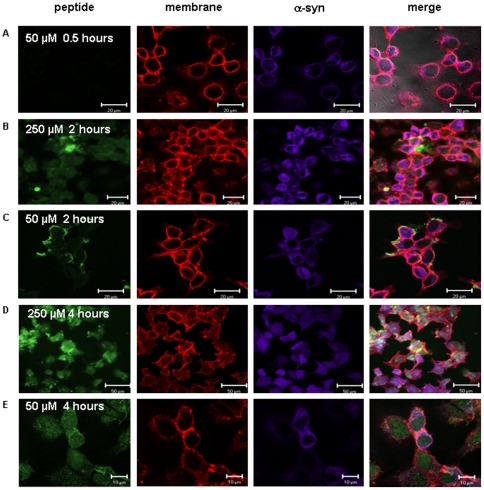
Figure 7. Internalization of retro-inverso β-syn 36 into the cells.
Differentiated SH-SY5Y cells over expressing wild type α-syn were incubated with 50 µM and 250 µM of FITC-conjugated retro-inverso β-syn 36 peptide for periods of half an hour, two hours and four hours. After fixation, the presence of the FITC-conjugated peptide (green) was monitored inside the cells. Cellular α-syn was detected using cy5-conjugated antibody (purple) and the cell membrane was marked with Phalloidin (red). (A) After 30 minutes at 37°C there was no peptide staining (green). (B–C) After two hours of incubation, no or little amount of peptide was detected inside the cells (green). (D–E) After 4 hours of incubation, the peptide was clearly detected inside the cells. Peptide localization was visualized using an LSM-510 Zeiss confocal microscope.

