Figure 5.
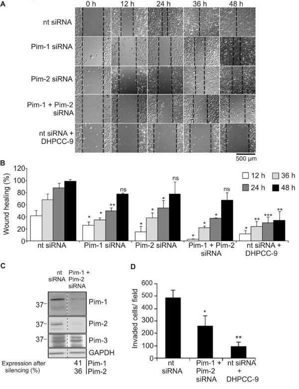
Silencing and inhibition of Pim kinases both repress migration and invasion of prostate cancer cells. PC-3 cells were transfected with 100 nM non-targeting (nt) control siRNA or siRNA oligonucleotides targeting pim genes, and incubated overnight. (A) For wound healing assays, transfected cells were moved onto 24-well plates and allowed to attach for 24 h, after which DMSO or 10 μM DHPCC-9 was added and scratch wounds were made with a sterile 200 μl pipette tip. Representative pictures were taken at indicated time-points and analysed. (B) Graph represents means of three independent experiments with duplicate samples. (C) Efficiency of Pim kinase silencing was determined by Western blotting. GAPDH staining was used as a loading control. Non-parallel lanes from a single electrophoresis gel are separated by dash lines. (D) For invasion assays, transfected cells were placed in invasion inserts together with either DMSO or 10 μM DHPCC-9. MG-63-conditioned medium was used as a chemoattractant to induce invasion. Cells were incubated for 72 h, after which insert membranes were fixed and stained. Invasion assays were repeated for three times and invaded cells were counted from each sample from 10 representative fields. Shown are means of duplicate samples from one representative experiment.

